Image One: Canine Maxillary X-ray
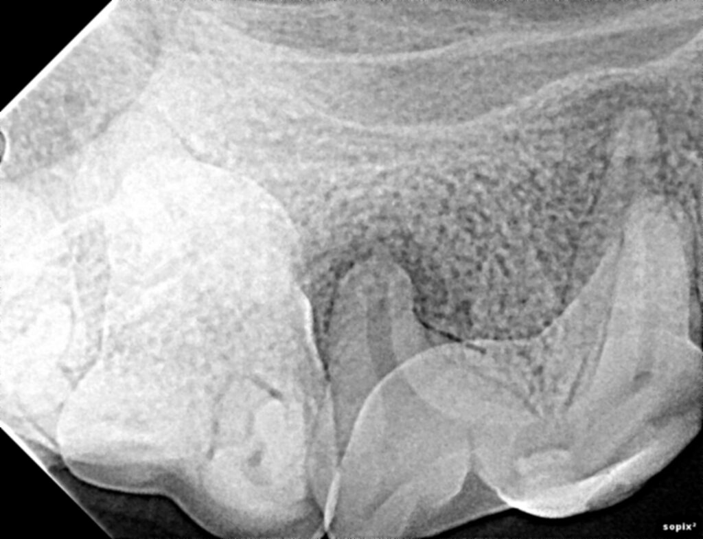
Comparing the two images DAVID has highlighted 4 anomalies. Three pathological and one non pathological.
Image One: DAVID analysis
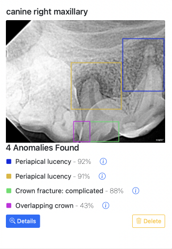
Marked in green is a complicated crown fracture. that is a crown fracture where the pulp cavity has been exposed.Marked in Blue and Yellow David has also detected the periodical licences around the roots of the last pre molar.
Marked in purple is an overlapping crown. Although non pathological it is common for inexperienced people to think this is problematic.
Image Two: Feline rostral Maxillary
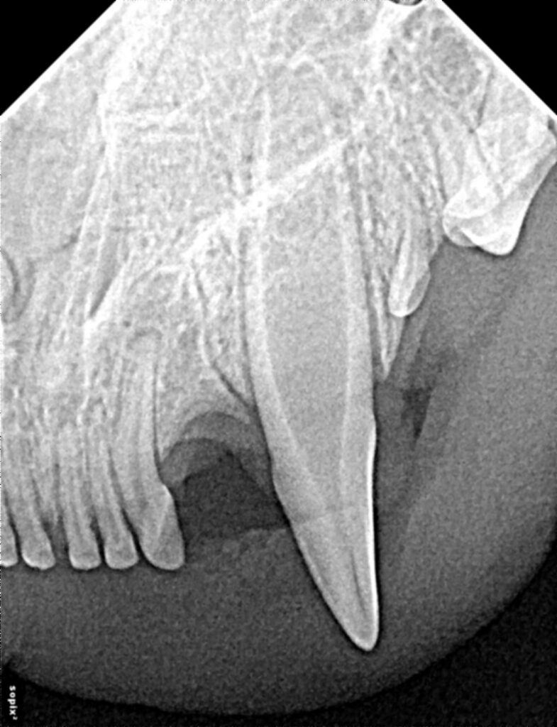
Image Two: DAVID Analysis
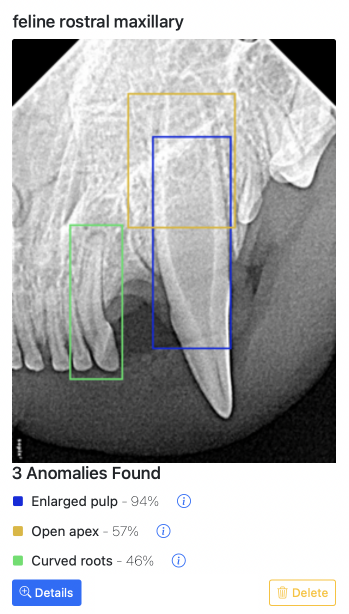
Comparing the two images DAVID has highlighted 3 anomalies. All three are non pathological.
Marked in Blue is the enlarged pulp of the canine tooth. This can be pathological. However in this case DAVID has correctly identified that it is normal as the patient is an immature feline. Marked in Yellow is the open apex of the same canine tooth. Again in this typical of an immature patient.
Marked in Green is the curved root of the third incisor. Although non pathological curved roots can complicate dental procedures so DAVID draws your attention to it.
Image Three: Photograph
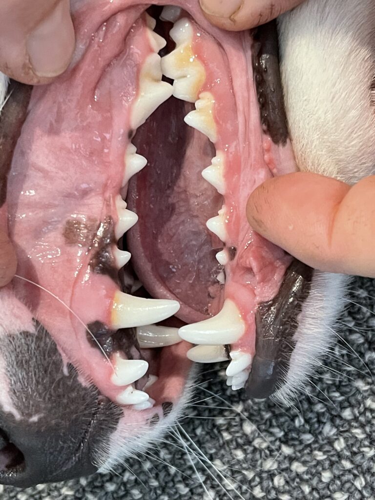
Comparing the two images DAVID has highlighted 17 anomalies all pathological from an image taken on a cell phone.
DAVID has highlighted multiple instances of gingivitis. Gingivitis is the first stage of periodontal disease and radiographs are indicated to assess the degree of disease.
Calculus has also been highlighted. The patient is a juvenile blue border collie and time was allocated for a full mouth series of radiographs and prophylaxis to clean the teeth.
The image with marked pathology was instramental in the owner accepting the recommended treatment plan.
Image Three: DAVID Analysis
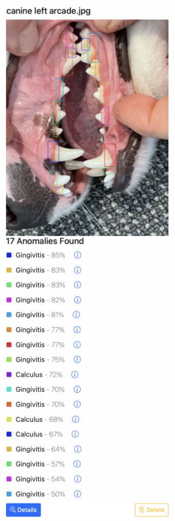
The DAVID images shown are the instant results. More detailed views and pathology and treatment options are available in the system.